


Brain Scans: More than Black and White
Six CAN-BIND Community Advisory Committee (CAC) teleconference meetings are held each year. During these sessions, a CAN-BIND research scientist is invited to give a brief talk about their field of research.
In a meeting held earlier this year, Dr. Stefanie Hassel, Research Manager for CAN-BIND’s Neuroimaging Platform, provided an overview of the importance of neuroimaging and the role her team plays in ensuring that the research CAN-BIND conducts is consistent and reliable. Learn more about Dr. Hassel’s talk below.
Why is neuroimaging important in depression research?
Depression is a serious illness that can affect brain structure, function and chemical balance, and behaviour. Researchers such as Dr. Mary Phillips (University of Pittsburgh) have developed brain models to help us better understand and compare structural differences and brain activity between people with and without mood disorders such as depression. Models like these allow us to use neuroimaging to identify and study brain areas affected by mood disorders, including:
- The limbic system. Tucked in behind the ear, areas of the limbic system are involved in emotional regulation (e.g. feeling happy, fearful or disgusted). In people with depression, some areas of the limbic system, such as the amygdala, have been found to be overly active.
- Areas of the prefrontal cortex (PFC). Located at the front of the head, parts of the PFC are involved in cognitive control. In people with depression, activity in these areas may not be properly regulated.
In addition to collecting data from biological samples (e.g. blood or urine) and questionnaires in research studies, neuroimaging allows us to collect different pieces of information needed to find biological markers or “biomarkers” that will help physicians to predict the most effective treatment plans for patients living with depression.
What neuroimaging techniques are used in CAN-BIND?
In CAN-BIND, the imaging techniques used are called structural magnetic resonance imaging (MRI), functional MRI (fMRI) and diffusion tensor imaging (DTI). fMRI is used to look at functional changes in the brain based on changes in blood flow in different brain areas. There is higher blood flow in more active brain areas and lower blood flow in less active brain areas. For example, when completing a memory task, areas of the brain involved in memory, such as the prefrontal regions, show differences in blood flow. In comparison, MRI and DTI are used to look at the brain’s structure. DTI looks at how well information travels through tracts (bundles of axons that connect different brain areas to each other). More information can be found here.
What does the CAN-BIND Neuroimaging Platform do?
As a team that works with many different research sites and collaborators across Canada, the neuroimaging team has multiple responsibilities.
One main role is working towards ensuring that all data are standardized. The team is in charge of creating and maintaining the neuroimaging Standard Operating Procedures (SOPs). SOPs are step-by-step written guidelines that are used across research sites to ensure research is carried out in a consistent manner. For neuroimaging, this involves setting up the MRI scanners in a specific way to scan study participants’ brains. This helps to ensure that the data collected are comparable and reliable and that the study can be replicated. SOPs are shared with all study investigators, coordinators, research students and neuroimaging technologists. The neuroimaging team is currently in the process of publishing an MRI protocol paper to help other researchers better understand how to standardize data.
The team is also in charge of assessing the usability of CAN-BIND study data (quality control) and analysing it.
Assessing the data’s usability ensures data are of the highest quality and results are representative of changes in brain structure or brain function and not due to an artefact.
Assessing the data’s usability ensures data are of the highest quality and results are representative of changes in brain structure or brain function and not due to an artefact. Artefacts are called “noise” in the data. How long these steps take can vary depending on the complexity and amount of data collected (usually a few months). Data quality control of the MRI, fMRI and DTI data from the CAN-BIND-1 Antidepressant Medication study has been completed. The analysis of these data is on-going.
After Dr. Hassel provided an overview of the role of CAN-BIND’s Neuroimaging Platform, the Community Advisory Committee (CAC) asked a few questions.
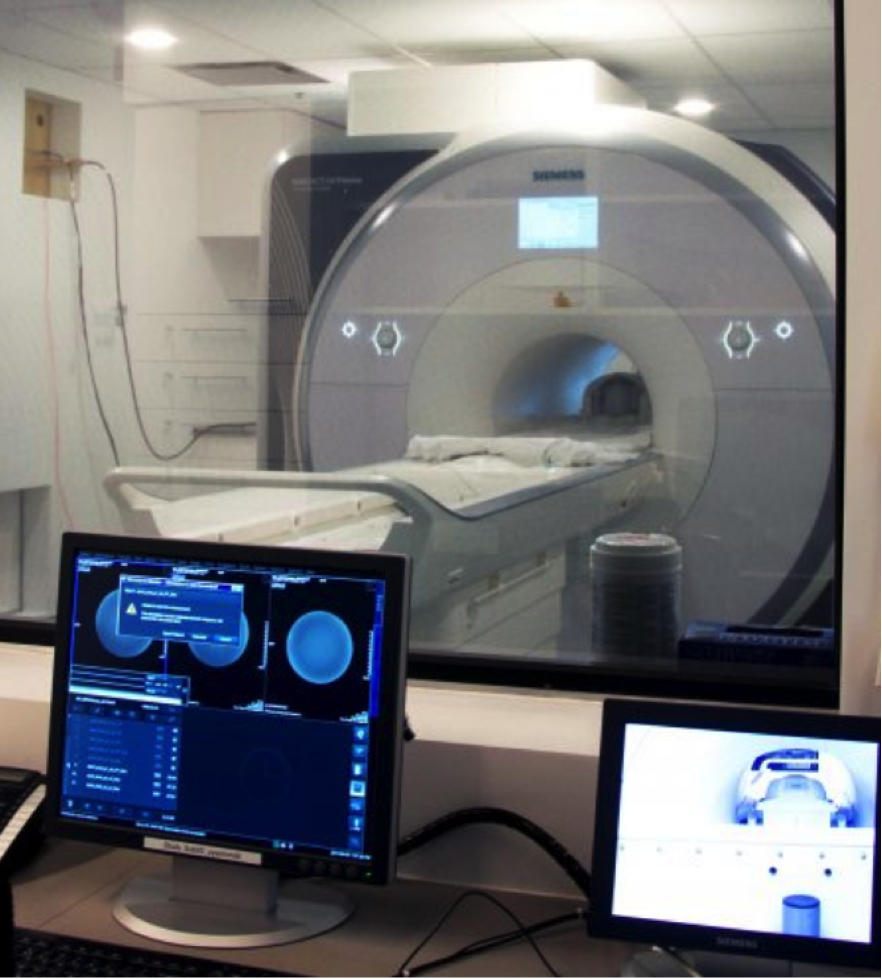
How long does a person stay in a brain scanner for?
It depends on what you are interested in exploring. To look at brain structure only, scan sequences can be as short as a few minutes (<10 minutes). For DTI and for fMRI, sequences tend to be a little longer; some tasks can last 10 minutes. For CAN-BIND, a typical scan is about 75 minutes because we explore all three different ways of looking at the brain. 75 minutes is considered a rather long time because one has to be very still during the scan, and because an MRI scanner is quite a noisy environment. Due to the nature of data collection, you need a lot of repeated measurements, which contributes to the longer amount of time.
How many times have you (Dr. Hassel) been scanned?
At least 10 times. If I ask a participant to do something, it’s important to me that I’ve experienced it myself. I have also tried transcranial magnetic stimulation (TMS). I strive to fully understand what is asked of participants, so that I can better explain the experience to individuals who are interested in joining a study, but have questions about the study protocol.
With the kind of scans used in CAN-BIND, can you see inflammation?
You cannot see inflammation with the brain imaging techniques CAN-BIND uses. We measure blood flow via fMRI and the integrity of tracts in the brain via DTI.
Dr. Sagar Parikh, Co-chair of the CAC, mentioned that there are other techniques, such as positron emission tomography (PET) imaging, which to some extent can detect brain inflammation.
Do all people who have depression have a similar fMRI image?
When analysing neuroimaging data, you have to look at groups of individuals to derive meaning from the results. On a group-level, we can say a particular group shows increased or decreased activation in a given, or specific, brain area. But for an individual, the pattern can be different.
There are definitely patterns of brain activation that seem to be specific to people who have depression compared to people who do not. The differences between individuals is something CAN-BIND is trying to identify and address.
Dr. Hassel concluded the talk by asking the CAC team for their feedback.
How can researchers make participating in a research study a more informative experience?
Suggestions included:
- Provide information (e.g. a leaflet) on what each scanning machine does and what it’s like being in a scanner.
- Provide opportunities for groups of potentially interested study participants to observe part of the (brain scan) process to see what it would be like.
- Often in clinical research, participants would love to hear about the results. Perhaps create a knowledge translation process to share the neuroimaging information (through newsletters or presentations). Reaching out to those who have taken the time to participate would be beneficial.
Do you have any suggestions for how researchers can make a neuroimaging experience more informative for research participants? Please email canbind@smh.ca with your ideas.
Further reading
Dr. Hassel provided the links (click on titles below) for those interested in learning more about fMRI and DTI.
- What is fMRI? By UC San Diego School of Medicine
- Brain Scans: Technologies That Peer Inside Your Head By Brainfacts.org
Have you met our Community Advisory Committee (CAC) members? Learn more about the CAN-BIND CAC, what members do, and read our collection of member spotlights on our website.
Authors: Anum Shivji Arwani, MSc and Janice Pong, MSc, Centre for Depression and Suicide Studies
Editors: Dr. Stefanie Hassel, PhD, Cumming School of Medicine, University of Calgary and Susan Rotzinger, PhD, CAN-BIND